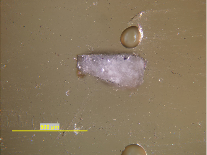
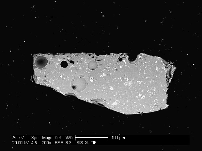
Figure 25a-b. Cross-section of light blue glaze on the plate. Blue is at the top edge of the sample in the optical microscope image (left). The extent of its penetration into the white glaze can be tracked through precipitates that appear dark in the backscattered electron image (right).